生物医学电镜研究网站 Bio-Medical EM Research Website
在充分利用电子显微镜卓越空间分辨率的同时,我们将加速以iPS细胞为首的再生医学研究,构建服务于其临床应用的《再生器官品质电镜评估系统》,并推出助力最新基因组编辑技术应用研究的《细胞形态/组织构建电镜评估系统》。
While utilizing the excellent spatial resolution of electron microscopes, we will accelerate research on regenerative medicine, including iPS cells, and build a "Electron Microscope Evaluation System for Regenerative Organ Quality" that contributes to its clinical application, and provide a "Electron Microscope Evaluation System for Cell Morphology/Tissue Construction" that contributes to applied research on the latest genome editing technology.
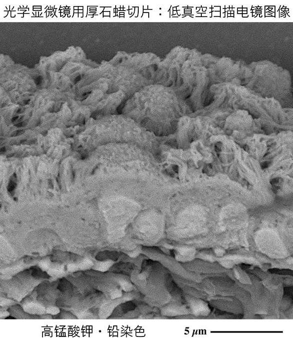
我们开发了一种基于高锰酸钾氧化反应的光学显微镜用石蜡切片电子染色法,可用于低真空扫描电子显微镜下的立体微观结构解析。
Development of a new a new metal staining method by potassium permanganate oxidation that allows uranium-free imaging of cell/tissue architectures in low-vacuum scanning electron microscopy
我们开发了一种突破性的“基于高锰酸钾氧化反应的电子染色法”,无需使用19世纪60年代开发的传统方法中所需的醋酸铀(国际管制物质),即可通过低真空扫描电子显微镜对光学显微镜用石蜡切片进行立体微观结构解析。
We have developed an epoch-making "uranium-free" metal staining method by potassium permanganate oxidation that allows uranium-free three-dimensional imaging of cell/tissue architectures in paraffin section by low-vacuum scanning electron microscopy
Sawaguchi, A. et al. npj Imaging 2, 40 (2024)
由日本科学技术振兴机构(JST)在《科学日本》介绍
由日本科学技术振兴机构(JST)在《客观日本》介绍
利用台式低真空扫描电子显微镜的优势和特性
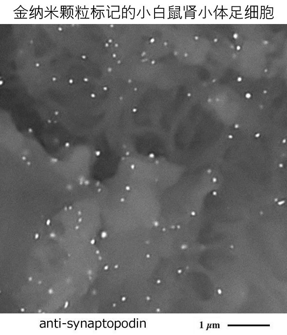
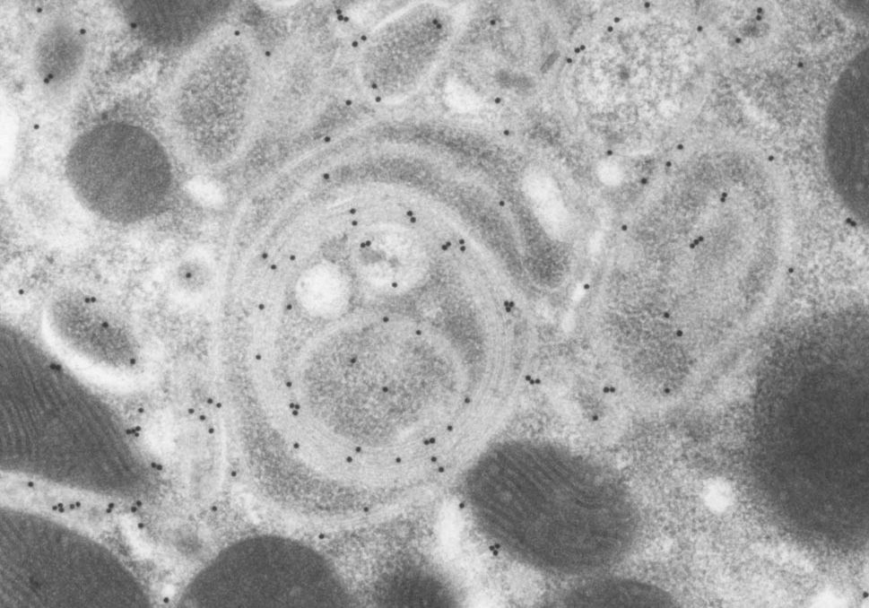
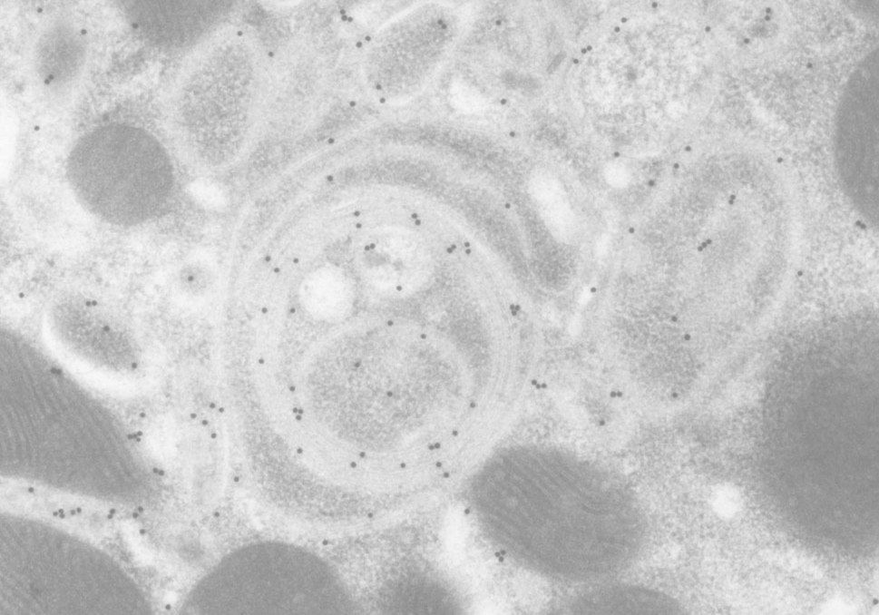
开发出一种可视化生物物质定位的全新金纳米颗粒标记法
Development of a new gold nanoparticle labeling method for visualizing the localization of biological materials by utilizing the advantages and properties of desktop low-vacuum scanning electron microscopy
我们开发了一种新型金纳米颗粒标记法,该方法基于酶标抗体法免疫组化染色的石蜡切片,通过氯化金处理原位生长金纳米颗粒,从而轻松实现目标生物物质的定位可视化。
We have developed a new gold nanoparticle labeling method that allows easy visualization of the localization of target biological material by growing gold nanoparticles formed by treating with gold chloride on paraffin sections stained with immunohistochemistry using an enzyme-antibody method.
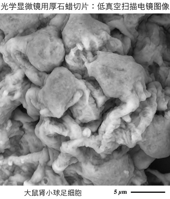
利用台式低真空扫描电子显微镜优势和特性的光学显微镜用
厚石蜡切片:高分辨率立体构建解析法的开发
Development of a high-resolution stereoscopic analysis method for thick paraffin sections by taking advantage of the advantages and characteristics of a desktop low-vacuum scanning electron microscope
我们开发了一种新型分析方法:充分发挥台式低真空扫描电镜的优势与特性,可对厚度为20-30微米的石蜡切片进行三维成像,捕捉构成生物体的组织与细胞结构,且该设备无需对切片进行抗荷电喷镀即可观察整张玻片。
We have developed a new analysis method that captures the tissues and cells that make up living organisms in three dimensions by taking advantage of the advantages and characteristics of a desktop low-vacuum scanning electron microscope, which can observe entire glass slides without applying antistatic treatment to paraffin sections sliced to a thickness of 20 to 30 μm.
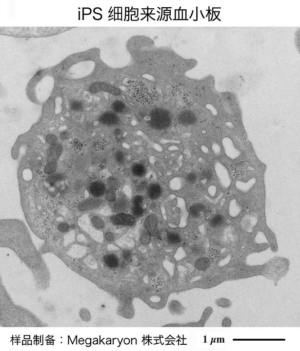
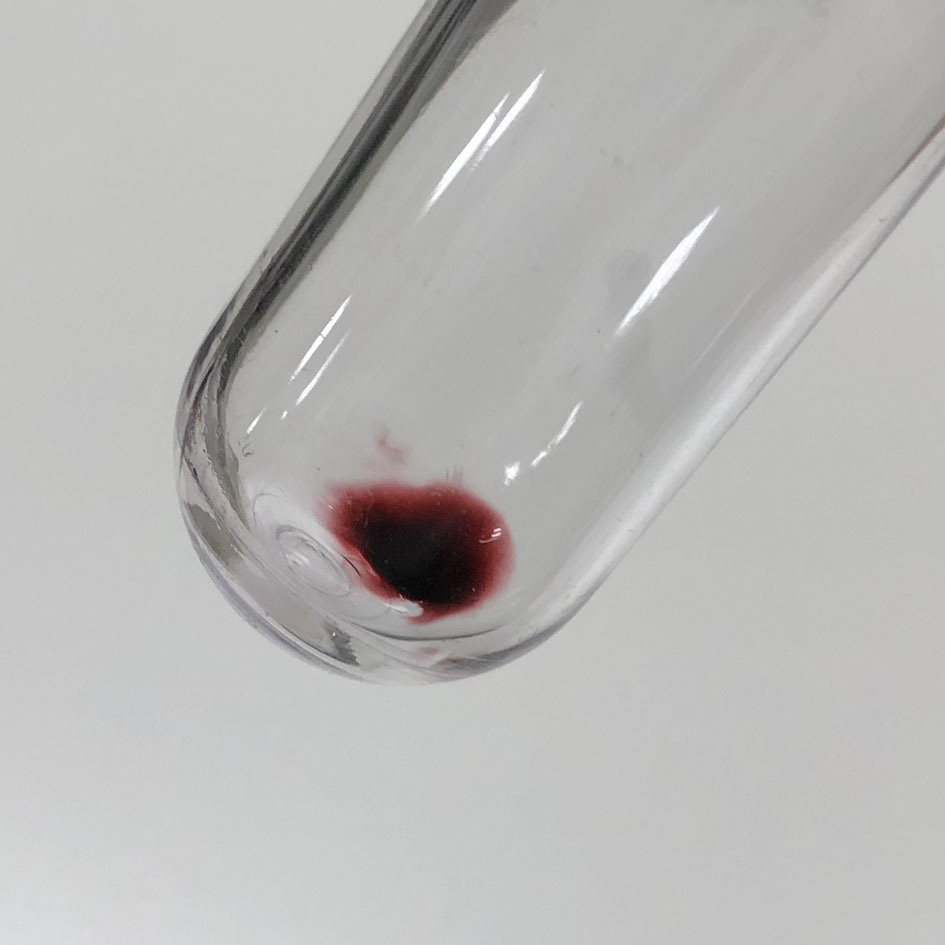
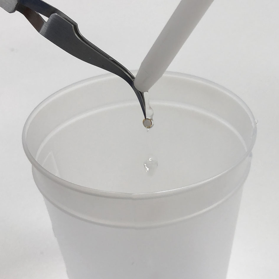
服务于iPS细胞来源血小板制剂开发与临床应用的电子显微镜解析
Electron Microscopical Evaluation of iPS Cell-Derived Human Blood Platelets for Clinical Transfusion
作为京都大学iPS细胞研究所(CiRA)江藤浩之教授实验室、Megakaryon株式会社以及日立高新技术株式会社(Hitachi High-Tech)的产学研合作项目,我们正在开展血小板及其前体细胞巨核细胞的超微形态分析,旨在能够早日实现iPS细胞来源血小板制剂的开发与临床应用。
As an industry-academia collaboration project with Prof. Koji Eto's lab at the Center for iPS Cell Research and Application (CiRA) in Kyoto University, Megakaryon Co., Ltd., and Hitachi High-Tech, Ltd., we are conducting ultra-micromorphological analysis of platelets and their progenitor cells, megakaryocytes, with a view to the early realization of iPS cell-derived platelet products and clinical applications.
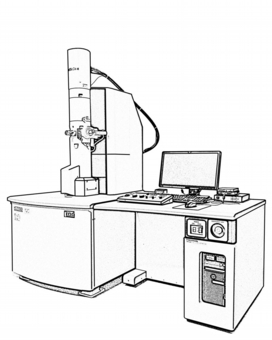
台式低真空扫描电子显微镜
Desktop Low-Vacuum Scanning Electron Microscopy
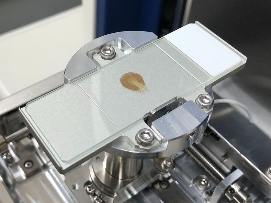
可对光学显微镜用石蜡切片连同载玻片进行高精细解析
Paraffin sections are analyzed in high definition on glass slides
详情请参阅此处
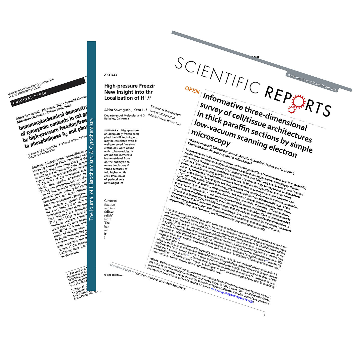
【参考文献列表】
低真空扫描电子显微镜:石蜡切片观察
Literature on Low-vacuum SEM Imaging of Paraffin sections
石蜡切片观察相关参考文献列表
Bibliography on paraffin section observation by Low-vacuum SEM
前往参考文献列表
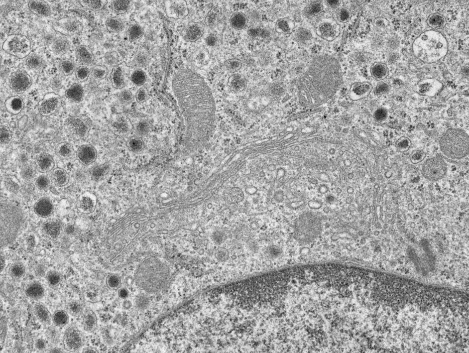
透射电子显微镜
TEM:Transmission Electron Microscopy
以最高水平的空间分辨率实现细胞内部的微观结构解析
Analysis of the fine structure of cells with high resolution
详情请参阅此处
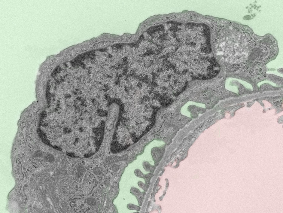
附读彩图指南,通俗易懂,秒懂图像!
电子显微镜图像集
Electron Micrograph Gallery:Easy-to-understand, image-reading color guide
展现器官再生研究所追求的精密生命设计图
Blueprint for Organ Regeneration Research
请欣赏高质量图像集
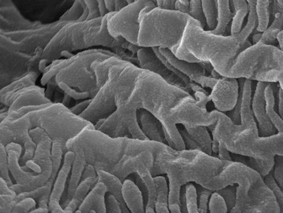
扫描电子显微镜
SEM:Scanning Electron Microscopy
立体、高倍放大观察样品表面,观察微观结构
Scanning over the surface of a sample in three dimensions at high magnification
详情请参阅此处
|
|
|
|
厚石蜡切片 高分辨立体图像集 |
宫崎大学前沿科学综合研究中心 生物成像实验室 |
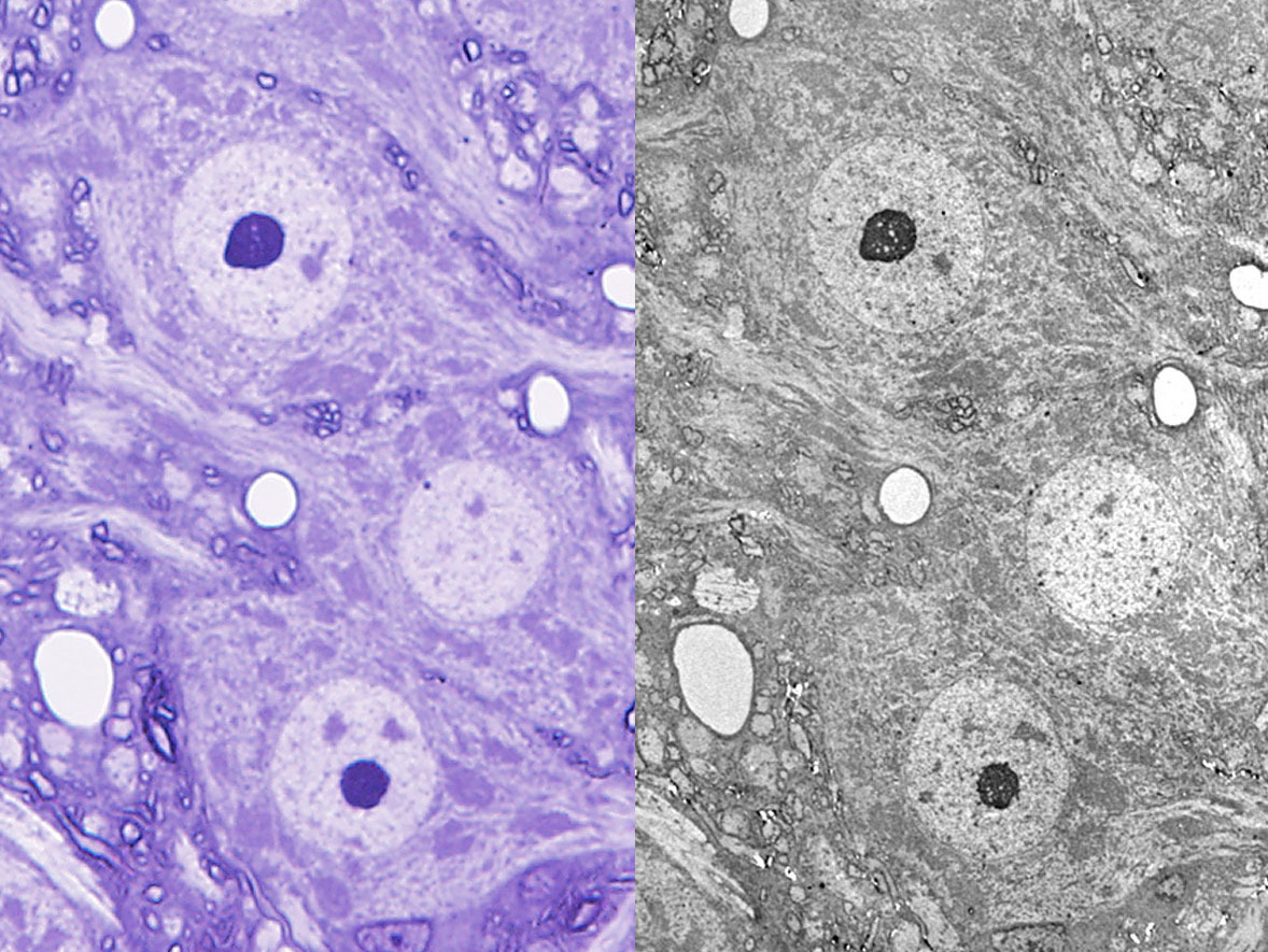
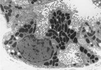
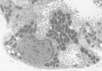
CLEM:光电关联显微技术 (Correlative Light and Electron Microscopy)
将光学显微镜筛选观察获得的最佳视野,定位至同一区域进行电镜观察!
在形态学分析中,建议先利用光学显微镜掌握观察对象的整体结构,然后再通过电子显微镜观察其细节。此外,在学术论文中,若能同时展示同一视野的光镜和电镜图像,将更具说服力。以下概述了光电关联显微技术(CLEM:Correlative Light and Electron Microscopy),即对甲苯胺蓝染色的超薄切片进行光镜观察与拍摄,再经树脂包埋后进行电镜观察。
In morphological analysis, it is recommended to first grasp the overall picture of the object to be observed at the light microscope level, and then observe the details at the electron microscope level. In addition, in academic papers, it will be more persuasive if electron microscope images with the same field of view can be presented alongside light microscopic images. This section provides an overview of Correlative Light and Electron Microscopy (CLEM), in which quasi-ultrathin sections stained with toluidine blue staining are observed and photographed by light microscopy, reembedded in resin, and electron microscopy is performed.
|
|
|
RESEARCH 研 究
利用电子显微镜卓越的空间分辨率,探索美丽的“生命形态”
We are exploring the beautiful "form of life" by taking advantages of the excellent spatial resolution of electron microscope
|
|
|
|
|
CONTACT US
Department of Anatomy, Ultrastructural Cell Biology
Faculty of Medicine, University of Miyazaki
Miyazaki 889-1692, Japan
TEL +81-985-85-1784 FAX +81-985-85-8406