【 英文総説 】
低真空走査電子顕微鏡の医学生物学研究への応用
Sawaguchi, A.: Low-vacuum scanning electron microscopy for informative three-dimensional imaging of cell/tissue architectures and biomedical target localization.
Microscopy, Feb 19: dfag012, 2026 Open Access
DOI: 10.1093/jmicro/dfag012
【 英文原著 】
ウラン化合物を必要としない画期的な過マンガン酸カリウム電子染色法の開発
Sawaguchi, A., Kamimura, T., Kitagawa, K., et al.: KMnO4/Pb staining allows uranium free imaging of tissue architectures in low vacuum scanning electron microscopy.
npj Imaging 2:40, 2024 Open Access
DOI: s44303-024-00045-z
血管内皮glycocaryxの観察に基づく血管病理研究
Mori, K., Tomita, H., Kuno, M., et al.: Practical method for endothelial glycocalyx imaging in formalin-fixed, paraffin-embedded tissues in vascular pathology.
Microvasc Res. 165:104913, 2026 Open Access
DOI: 10.1016/j.mvr.2026.104913
ヘリコバクター・フェリスによる急性胃粘膜傷害の一例
Aman, M., Gi, T., Maekawa, K., Kimura, T., Sawaguchi, A., Yamashita, A.: Helicobacter felis‐Associated Acute Gastric Mucosal Lesion: Ultrastructure by Low‐Vacuum Scanning Electron Microscopy.
Pathol Int. 76(4): e70109, 2026
DOI: 10.1111/pin.70109
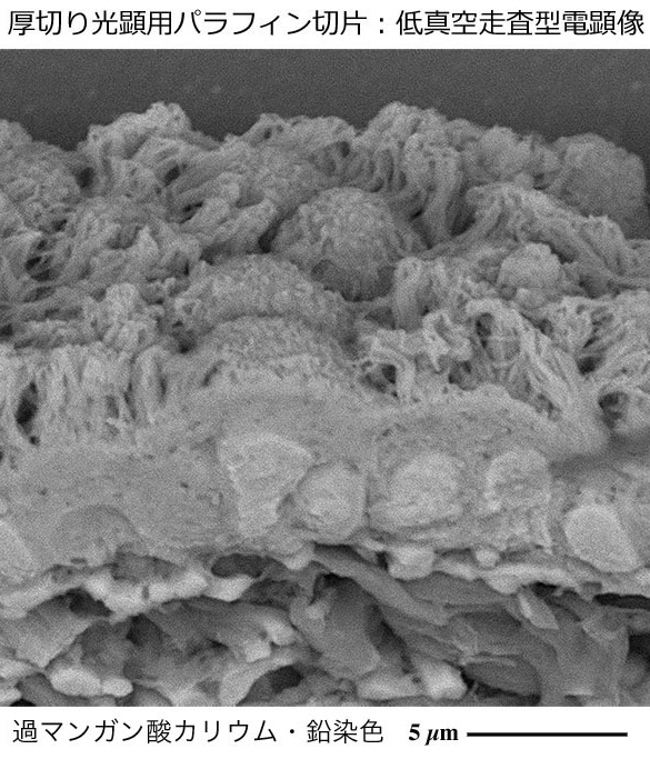
過マンガン酸カリウムの酸化反応を応用した光顕用パラフィン切片
電子染色法の開発と低真空走査電子顕微鏡による立体微細構造解析
KMnO4/Pb staining allows uranium free imaging of tissue architectures in low vacuum scanning electron microscopy.
1960年代に開発された従来法の酢酸ウラニウム(=国際規制物資)を必要としない画期的な「過マンガン酸カリウム酸化反応を応用した電子染色法」を開発し、低真空走査電子顕微鏡による光顕用パラフィン切片の立体微細構造解析を可能にしました。
ラット頭蓋骨欠損モデルにおけるバイオミネラリゼーションに関する多角的分析
Shimada, N., Hirata, A., Yamada, S., et al. Multimodal analysis of biomineralization within a collagen scaffolding in a rat calvarial defect model by using decalcified and undecalcified specimens
J Mater Sci Mater Med. 37:30, 2026 Open Access
DOI: 10.1007/s10856-026-07003-8
神経性やせ症患者に生じた腎尿細管結晶に関する症例報告
Zhu, Y., Kawanishi, K., Sawa, C., et al. Application of low-vacuum scanning electron microscopy and energy-dispersive X-ray spectrometry for detection of renal tubular crystals: a case of nephrocalcinosis in the setting of anorexia nervosa.
BMC Nephrology 26:352, 2025 Open Access
DOI: 10.1186/s12882-025-04287-w
深部静脈血栓症における第XI因子の局在と生理機能に関する研究
Oguri, N., Gi, T., Nakamura, E., et al. Factor XI localization in human deep venous thrombus and function of activated factor XI on venous thrombus formation and hemostasis.
Res Pract Thromb Haemost 9:e102720, 2025 Open Access
DOI: 10.1093/ehjcr/ytae196
ネイル・パテラ症候群患者で生じたアルポート症候群に近似した腎糸球体基底膜病変
Ito, H., Ishiyama, K., Hirose, T., et al. The first observation and diagnosis of nail-patella syndrome using LV-SEM: GBM abnormalities mimicking Alport syndrome.
Nephrology 30:e70062, 2025 Open Access
DOI: 10.1111/nep.70062
大腿動脈石灰化結節症病理組織の三次元的微細構造解析
Nishimura, M., Yano, M., Nishihira, K., et al. Three-dimensional fine structure of calcified nodules in the common femoral artery based on low-vacuum scanning electron microscopy.
Eur Heart J-Case Reports- 8:ytae196, 2024 Open Access
DOI: 10.1093/ehjcr/ytae196
ラット角膜アルカリ炎症モデルにおけるマクロファージ浸潤抑制に関する微細形態研究
Ikebukuro, T., Arima, T., Kasamatsu, M., et al. Disulfiram ophthalmic solution inhibited macrophage infiltration by uppressing Mamcrophage pseudopodia formation in a at corneal alkali burn model.
Int J Mol Sci. 24: 735, 2023 Open Access
DOI: 10.3390/ijms24010735
クロイツフェルトーヤコブ病に関する脳病理組織所見
Shijo, M., Yoshimura, M., Omae, T., et al. Altered properties of amyloidogenic prion protein in genetic Creutzfeldt-Jakob disease with PRNP V180I mutation in response to pentosan polysulfate.
Brain Pathol. 33(5): e13197, 2023 Open Access
DOI: 10.1111/bpa.13197
前眼房炎症におけるZinn小帯の組織所見
Takahashi, A., Arima, T., Toda, E., et al. A Novel Multi-Observation System to Study the Effects of Anterior Ocular Inflammation in Zinn's Zonule Using One Specimen.
Int J Mol Sci. 24(7): 6254, 2023 Open Access
DOI: 10.3390/ijms24076254
凍結固定法による筋膜の超微形態観察
Imazato, H., Takahashi, N., Hirakawa, Y., et al. Three-dimensional fine structures in deep fascia revealed by combined use of cryo-fixed histochemistry and low-vacuum scanning microscopy.
Sci Rep. 13(1): 6352, 2023 Open Access
DOI: 10.1038/s41598-023-33479-3
腎糸球体基底膜及び足細胞の微細構造変化
Lan, P., Kang, D., Mii, A., et al. Evaluation of ultrastructural alterations of glomerular basement membrane and podocytes in glomeruli by low-vacuum scanning electron microscopy.
Clin Exp Nephrol. 26(3):216-225, 2022 Open Access
DOI: 10.1007/s10157-021-02147-z
DAB標識免疫染色の電子染色増感法
Arai, Y., Takeuchi, K., Hatanaka, S., et al. Heavy Metal Enhancement Technique for Diaminobenzidine in Immunohistochemistry Enables Ultrastructural Observation by Low-vacuum Scanning Electron Microscopy.
J Histochem Cytochem. 70(6): 427-436, 2022 Free PMC article
DOI: 10.1369/00221554221102996
紫色採尿バッグ症候群の解析事例
Abe, M., Furuichi, M., Ishimitsu, T., et al. Analysis of purple urine bag syndrome by low vacuum scanning electron microscopy.
Med Mol Morphol. 55(2):123-130, 2022 Open Access
DOI: 10.1007/s00795-022-00313-0
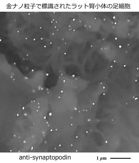
デスクトップ型低真空走査電子顕微鏡の利点・特性を活かした
生体物質の局在を可視化する新たな金ナノ粒子標識法の開発
In situ strategy for biomedical target localization via nanogold nucleation and secondary growth
酵素抗体法を用いて免疫組織化学染色された光顕用パラフィン切片に塩化金酸処理を施して形成された金ナノ粒子を成長させ、標的生体物質の局在を電子顕微鏡で容易に可視化する新たな金ナノ粒子標識法を開発しました。
新たな金ナノ粒子標識法の開発
Sawaguchi, A., Kamimura, T., Takahashi, N., et al. In situ strategy for biomedical target localization via nanogold nucleation and secondary growth.
Commun Biol. 4(1):710, 2021 Open Access
DOI: 10.1038/s42003-021-02246-3
ループス腎炎における病理組織所見
Yoshida, M. Hirashio, S., Doi, T., et al. Low-Vacuum Scanning Electron Microscopy to Assess Histopathological Resolution of Class V Lupus Nephritis: A Case Report.
Case Rep Nephrol Dial. 11(1): 36-47, 2021 Free PMC article
DOI: 10.1159/000509470
血管内皮glysocalyxのアルシアンブルー・銀増感染色
Mukai, S., Takaki, T., Nagumo, T., et al. Three-dimensional electron microscopy for endothelial glycocalyx observation using Alcian blue with silver enhancement.
Med Mol Morphol. 54(2):95-107, 2021 Open Access
DOI: 10.1007/s00795-020-00267-1
角膜創傷治癒と血管新生に関するパラフィン切片観察
Arima T., Uchiyama, M., Shimizu, A., et al. Observation of Corneal Wound Healing and Angiogenesis Using Low-Vacuum Scanning Electron Microscopy.
Transl Vis Sci Technol. 9(6): 14, 2020 Open Access
DOI: 10.1167/tvst.9.6.14
腎移植後の早期糸球体病変に関するパラフィン切片観察
Onishi, H., Oguchi, H., Shinoda, K., et al. Pathological Analysis of Early Transplant Glomerulopathy in Renal Allografts Using Low-Vacuum Scanning Electron Microscopy.
Nephron. 144 Suppl 1: 71-78, 2020 Free PMC article
DOI: 10.1159/000512136
ヒト腎移植後の抗体を介した拒絶反応の早期診断
Yokoyama, H., Okada, S., Yamada, Y., et al. Low-vacuum scanning electron microscopy may allow early diagnosis of human renal transplant antibody-mediated rejection.
Biomed Res. 41(2): 81-90, 2020 Free PMC article
DOI: 10.2220/biomedres.41.81
ラット角膜病変モデルにおいて水素がもたらす活性酸素分解酵素の活性化作用
Arima, T., Igarashi,T., Uchiyama, M., et al. Hydrogen promotes the activation of Cu, Zn superoxide dismutase in a rat corneal alkali-burn model.
Int J Ophthalmol. 13(8): 1173-1179, 2020 Open Access
DOI: 10.18240/ijo.2020.08.01
ナノスーツ法を応用したパラフィン切片観察
Kawasaki, H., Itoh, T., Takaku, Y., et al. The NanoSuit method: a novel histological approach for examining paraffin sections in a nondestructive manner by correlative light and electron microscopy.
Lab Invest. 100(1): 161-173, 2020 Open Access
DOI: 10.1038/s41374-019-0309-7
腎生検標本における糸球体及び基底膜の病理学的微細構造解析
Kajimoto, Y., Endo, Y., Terasaki, M., et al. Pathologic glomerular characteristics and glomerular basement membrane alterations in biopsy-proven thin basement membrane nephropathy.
Clin Experiment Nephrol. 23: 638-649, 2019
DOI: 10.1007/s10157-018-01687-1
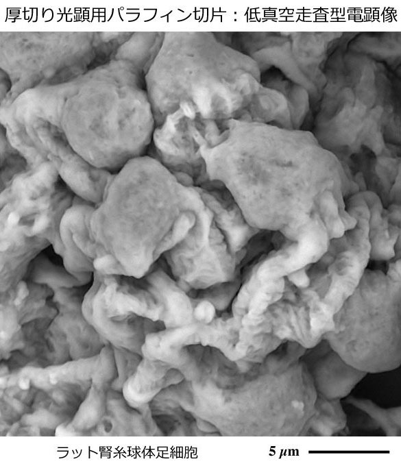
デスクトップ型低真空走査電子顕微鏡の利点・特性を活かした
厚切り光顕用パラフィン切片:高解像立体構築解析法の開発
Informative three-dimensional survey of cell/tissue architectures in thick paraffin sections
by simple low-vacuum scanning electron microscopy
厚さ20〜30 µmに薄切した光顕用パラフィン切片に帯電防止処理を加えず、スライドガラスごと観察できるデスクトップ型低真空走査電子顕微鏡の利点・特性を活かし、生体を構成する組織や細胞を立体的に捉える新たな解析手法を開発しました。
厚切り光顕用パラフィン切片:高解像立体構築解析法の開発
Sawaguchi, A., Kamimura, T., Yamashita, A., Takahashi, N., Ichikawa, K., Aoyama, F., Asada, Y.: Informative three-dimensional survey of cell/tissue architectures in thick paraffin sections by simple low-vacuum scanning electron microscopy.
Sci Rep. 8(1): 7479, 2018 Open Access
DOI: 10.1038/s41598-018-25840-8
角膜病変の修復機転における血管内皮増殖因子抑制作用に関する研究
Arima, T., Uchiyama, M., Nakano, Y., et al. Peroxisome proliferator-activated receptor alpha agonist suppresses neovascularization by reducing both vascular endothelial growth factor and angiopoietin-2 in corneal alkali burn.
Sci Rep. 7: 17763, 2017 Open Access
DOI: 10.1038/s41598-017-18113-3
パパニコロー染色病理診断への応用
Yano, T., Soejima, Y., Sawabe, M. Application of low vacuum scanning electron microscopy for Papanicolaou-stained slides for cytopathology examinations.
Microscopy. 65: 269–273, 2016
https://doi.org/10.1093/jmicro/dfw005
IgA腎症における糸球体基底膜傷害の病理組織所見
Masuda, Y., Yamanaka, N., Ishikawa, A., et al. Glomerular basement membrane injuries in IgA nephropathy evaluated by double immunostaining for α5(IV) and α2(IV) chains of type IV collagen and low-vacuum scanning electron microscopy.
Clin Exp Nephrol. 19(3): 427-435, 2015
DOI: 10.1007/s10157-014-1008-8
小児ネフローゼ症候群病理組織診断への応用
Okada, S., Inaga, S., Kawabata, Y., et al. A novel approach to the histological diagnosis of pediatric nephrotic syndrome by low vacuum scanning electron microscopy.
Biomed Res. 35: 227–236, 2014 Free Article
https://doi.org/10.2220/biomedres.35.227
アルポート症候群病理組織診断への応用
Okada, S., Inaga, S., Kitamoto, K., et al. Morphological diagnosis of Alport syndrome and thin basement membrane nephropathy by low vacuum scanning electron microscopy.
Biomed Res. 35(5): 345-350, 2014 Free Article
DOI: 10.2220/biomedres.35.345
腎生検病理組織診断への応用
Miyazaki, H., Uozaki, H., Tojo, A., et al. Application of low-vacuum scanning electron microscopy for renal biopsy specimens.
Pathol Res Pract. 208(9): 503-509, 2012
DOI: 10.1016/j.prp.2012.05.006
腎生検病理組織の三次元的微細構造解析
Inaga, S., Kato, M., Hirashima, S., et al. Rapid three-dimensional analysis of renal biopsy sections by low vacuum scanning electron microscopy.
Arch Histol Cytol. 73(3): 113-125, 2011 Free Article
DOI: 10.1679/aohc.73.113
白金ブルー染色法によるパラフィン切片観察
Inaga, S., Hirashima, S., Tanaka, K., et al. Low vacuum scanning electron microscopy for paraffin sections utilizing the differential stainability of cells and tissues with platinum blue.
Arch Histol Cytol. 72: 101–106, 2009 Free Article
https://doi.org/10.1679/aohc.72.101